
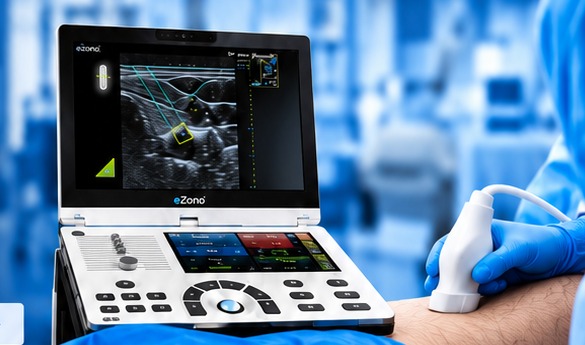
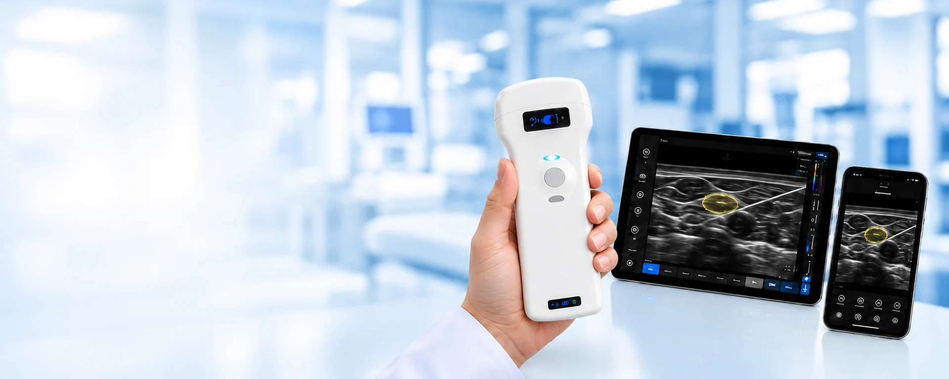

SonoStar Wireless Ultrasound
Portable wireless ultrasound for a wide range of clinical applications.
SonoStar
Wireless Wi-Fi operationSmall & lightweightHigh-quality imagingiOS / Android / WindowsEasy-to-use softwareTraining & telemedicine
Innovation in ultrasound imaging
Portable wireless ultrasound with an emphasis on ease of use and clinical flexibility.
- Quality technology in a compact format
- Wireless operation without cables
- Rechargeable operation
- Multiple probe families
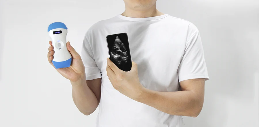
Flexibility for every clinical need
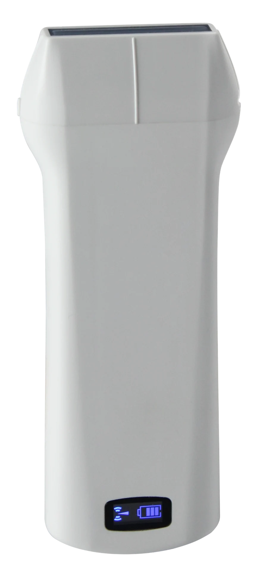 Linear
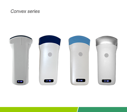
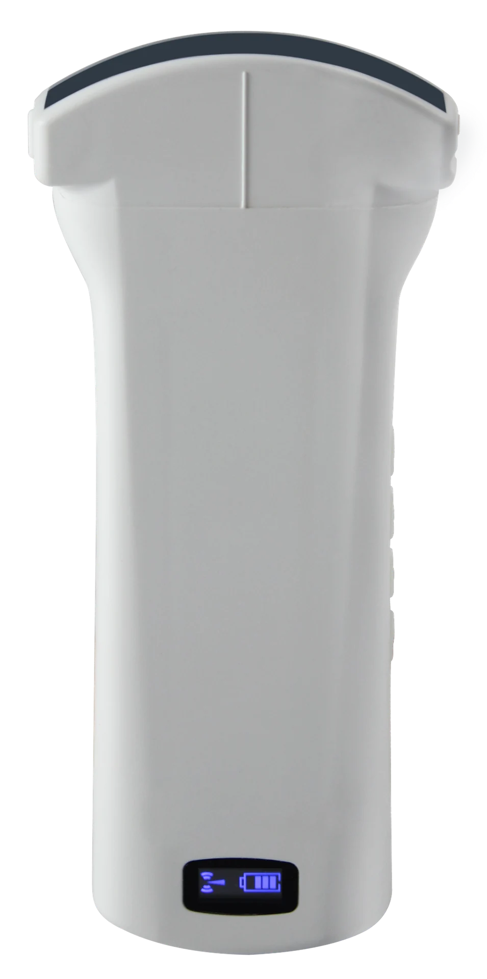 Convex
 Transvaginal
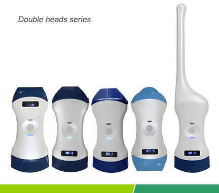
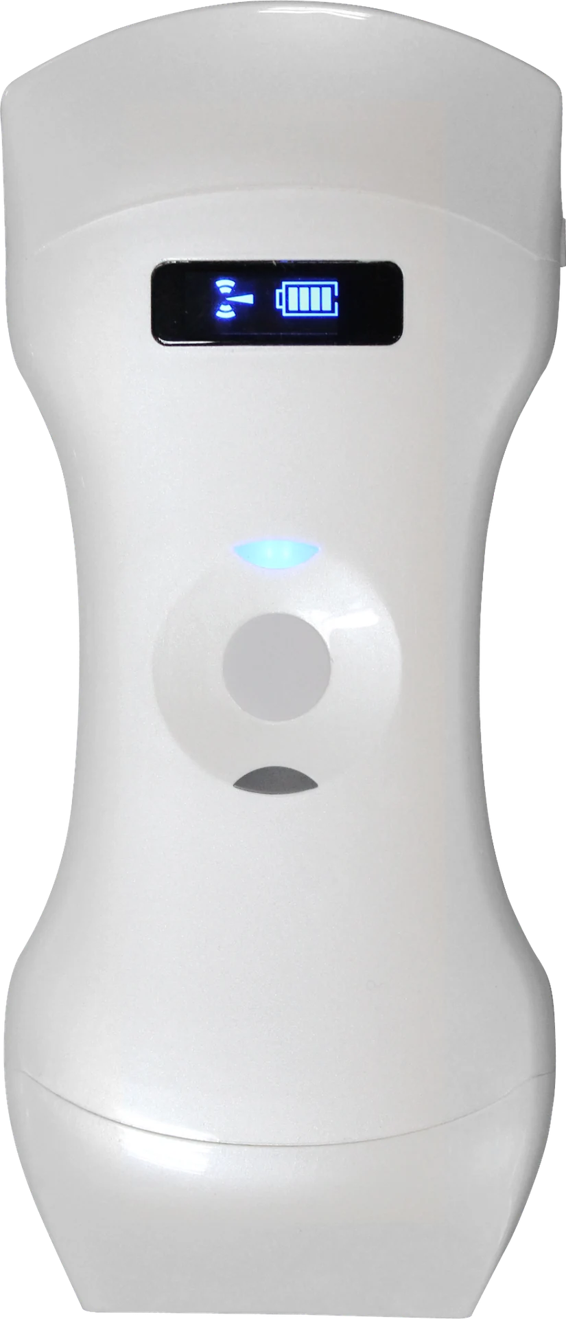 Dual / Triple heads
DUAL / TRIPLE HEADS
Linear • Convex • Sector
Combined heads for greater flexibility with one portable device.
General medicineEmergencyCardiologyAngiologyMSKAnaesthesiology
Imaging modes
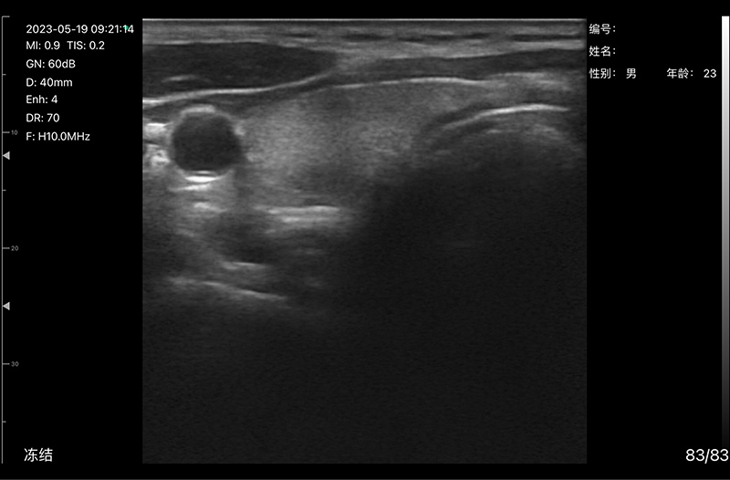
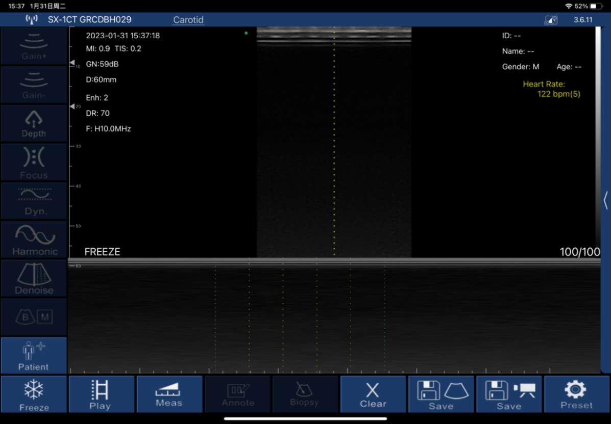
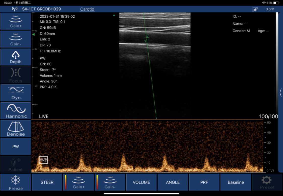
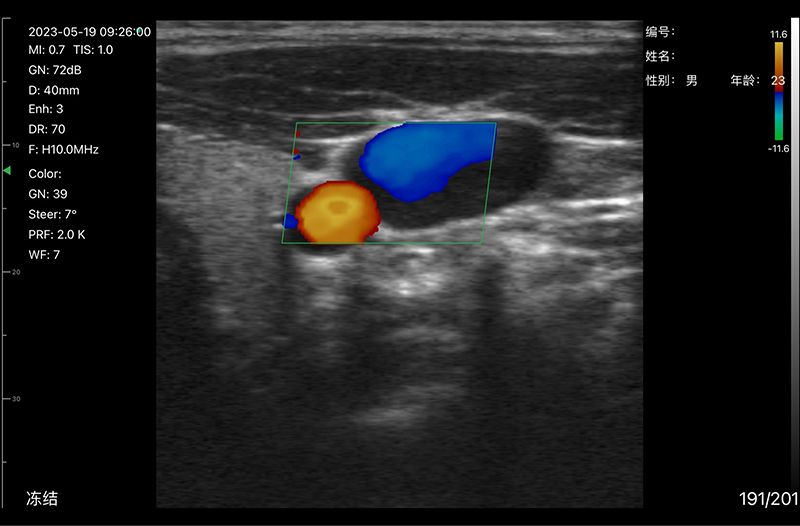
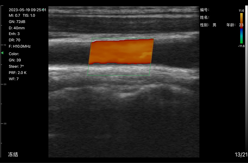
Example SonoStar images and interfaces. Availability depends on probe model. Select an image to view it in full.
VISION8 S High-frequency probe
Dermatology · Plastic surgery
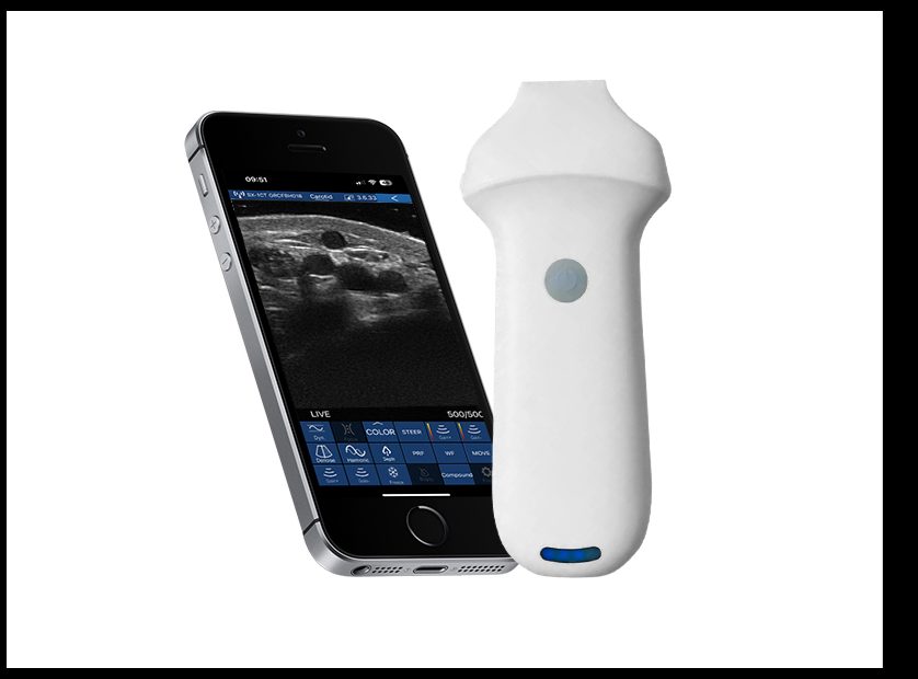
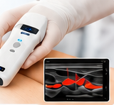
Dedicated 18/22 MHz head for dermatology, plastic surgery, small organs, peripheral nerves and microvascular assessment.
- 18 / 22 MHz
- 192 elements / 64 channels
- Superficial tissues and small organs
- Peripheral nerves and microvascular assessment
- Dermatology & plastic surgery
Imaging applications


The SonoStar family covers applications from general medicine and emergency care to cardiology, angiology, obstetrics/gynaecology, MSK and anaesthesiology.
General MedicineEmergency / AmbulanceICUCardiologyAngiologyObstetrics / GynaecologyMSKAnaesthesiology
Features
- Advanced technology at a highly economical price.
- High-quality digital imaging.
- Small, compact design for easy transport.
- Wireless operation without a cable.
- Compatibility with iOS and Android.
- Fluid-resistant design that supports cleaning and disinfection according to the instructions for use.
- Quality, durable housing.
- Connection and battery-status display.
- Support for remote diagnosis and image sharing.
- Intelligent tablet, mobile and PC software with imaging, storage, communication and printing functions.
- Wireless charging capability, depending on the model.
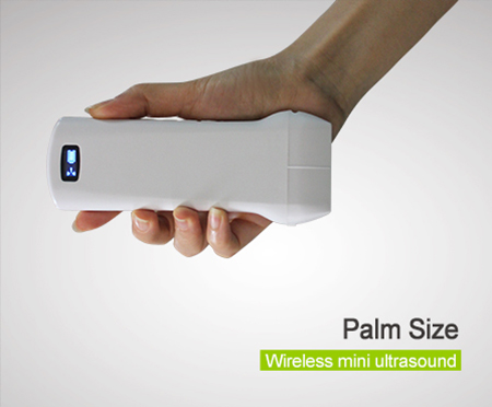
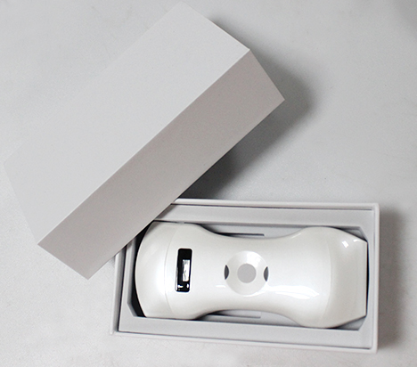
Flexibility
Wireless ultrasound probes are designed for a broad range of diagnostic examinations, with options for different anatomical regions and clinical applications.
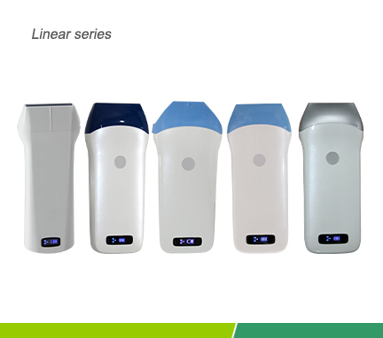
Single-head Linear, Convex, Phased Array, Microconvex and Transvaginal probes are available, alongside two-head solutions such as Convex + Linear and Convex + ICT.
Deep Blue & White Series offer colour Doppler imaging, while Blue & Grey Series offer monochrome B-mode imaging, depending on the model.
Imaging and budget options include 80 elements / 16 processing channels, 128 elements / 32 channels and 192 elements / 64 channels.
Uprobe L6C technical specifications
| Scanning type | Electronic array |
|---|---|
| Imaging modes | B, B/M, B+Color, B+PDI, B+PW |
| Probe elements | 192 |
| RF processing channels | 64 |
| Probe frequency | 7.5 / 10 MHz |
| Scanning depth | 20 / 40 / 60 / 100 mm |
| Probe width | 40 mm |
| Image processing | BGain, TGC, DYN, Focus, Depth, Harmonic, De-noise, Color Gain, Steer, PRF |
| CinePlay | Automatic and manual — 100 / 200 / 500 / 1000 frames |
| Biopsy guidance | Needle-guidance line for in-plane and out-of-plane approaches |
| Measurements | Length, Area, Angle, heart rate, Obstetrics |
| Image storage | JPG, AVI and DICOM |
| Frame rate | 18 frames / second |
| Battery | 3–5 hours of operation |
| Charging | USB or wireless charging — 2 hours |
| Dimensions | 156 × 60 × 20 mm |
| Weight | 220–250 g |
| Wi-Fi connection | 802.11g / 20 MHz / 5G / 450 Mbps |
| Operating system | iOS, Android and Windows |
These specifications apply to Uprobe L6C. Features of other probes vary by model.
Request a quote or demonstration
Tell us what you need for this product.
HEART MEDICAL
Contact →Presentation, training & support
Contact us for a product presentation, technical information and clinical support.