Negativation masking
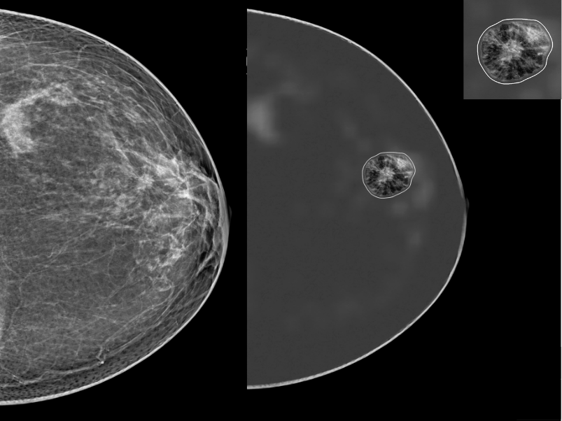
The optional “negativation” display masks the physiological background to make potentially suspicious areas stand out.


AI support for mammography reading
Hera-MI
 Breast-SlimView® · 2D / 3D
Hera-MI’s patented software provides a second look for radiologists, supporting the clinical assessment of 2D mammography and 3D tomosynthesis.
It highlights potentially suspicious areas, provides a risk score and estimates breast density using the BI-RADS 5th edition classification. Consistent review support helps radiologists focus on areas of interest during everyday reading.
The optional “negativation” display masks the physiological background to make potentially suspicious areas stand out.
Identification and characterisation of areas of interest, with a risk score for each area.
Comparative reading with assessment of breast density and risk evolution.
A structured summary for each breast, including density, risk score and detection localisation.
AI-based calculation supporting consistent classification according to BI-RADS 5th edition.
Projection of areas of interest onto an ultrasound pictogram to orient complementary examinations. The suite offers correlation capabilities across 2D/3D mammography, ultrasound and MRI, depending on the available modules.
Technical assessment and monitoring of acquisition quality for continuous practice improvement.
A summary of 2 to 4 synthetic 2D images per volume through the relevant module.
An example of the original mammographic image and a display highlighting an area of interest. Select the image to view it in full.
An AI tool for assessing and monitoring the quality of 2D and 3D mammographic images. It supports radiologists and radiographers in identifying opportunities for improvement and training needs.
Analysis of criteria based on the PGMI method.
Assessment of applied pressure in relation to breast type.
Information based on BI-RADS 5th edition.
Dose reporting and relevant acquisition data.
Select a thumbnail to open the corresponding full PDF. Identifying details have been removed from the sample headers.
Acquisition of examination data.
Automatic analysis within the local infrastructure.
DICOM results displayed within the radiologist’s environment.
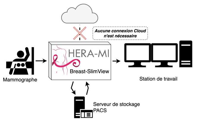
Processing integrates into the standard workflow without an additional initiation step by the radiologist or radiographer. It uses images already acquired and does not require additional radiation exposure. Final clinical assessment remains the physician’s responsibility.
Final requirements are sized according to mammography units, tomosynthesis, prior examinations and MTE modules.
Licence scope and additional modules are specified in the installation quotation.
Manufacturer: Hera-MI — Breast-SlimView® Suite
Tell us what you need for this product.
Contact us for a product presentation, technical information and clinical support.
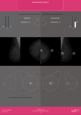 BSV — Localisation & prior comparison report
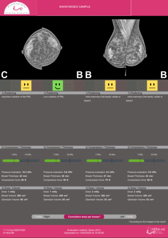 MTE — Image quality report